Showing posts with label human fibroblasts. Show all posts

Saturday, March 30, 2013

Allele Publishes mNeonGreen as the Brightest Monomeric Fluorescent Protein for Super-resolution Imaging

From AlleleNewsRelease: http://blog.allelebiotech.com/category/ips-and-other-stem-cells/page/2/

This week scientists from Allele Biotechnology and its partner non-profit research institute, the Scintillon Institute, present their latest fluorescent protein, mNeonGreen, in the journal Nature Methods (Nature Publishing Group). In the paper, entitled “A bright monomeric green fluorescent protein derived from Branchiostoma lanceolatum,” the scientists describe the development of the brightest monomeric fluorescent protein to date.
The scientific efforts to develop this novel fluorescent protein were led by Dr. Nathan Shaner, a leader in the field of fluorescent protein engineering. Fluorescent proteins are highly valuable research tools that allow the labeling and imaging of individual proteins within a 
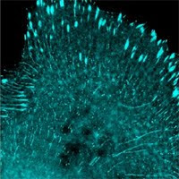
living cell, and tracking of their movements and localization in real time through a microscope. However, since the discovery of the original green fluorescent protein in 1993, imaging technology has advanced rapidly beyond the capability of most fluorescent proteins. The newly described fluorescent protein, mNeonGreen, allows researchers to take full advantage of modern super-resolution optical microscopy techniques that enable visualization of structures in living and fixed cells at much smaller scales than are possible using traditional optical microscopy. This improvement will lead to countless new insights into human health and a greater understanding of protein interactions at very small distance scales within living cells. According to Dr. Jiwu Wang, the CEO of Allele Biotechnology, “Super-resolution imaging will become the standard for publication in a short period of time, and mNeonGreen allows researchers to meet this standard while still being compatible with the equipment and methods they already use.”
Prominent researchers within the fluorescent protein field are touting mNeonGreen as a replacement for jellyfish-derived Aequorea GFP, one of the most commonly used fluorescent proteins today. According to lead researcher Dr. Nathan Shaner, “mNeonGreen can be directly substituted for other green fluorescent proteins such as EGFP without the need for any equipment changes,” making the upgrade an attractive prospect for many researchers.
Allele Biotechnology and Pharmaceuticals Inc. is a San Diego-based biotechnology company specializing in the fields of RNAi, stem cells, viral expression, camelid antibodies and fluorescent proteins. The company has co-developed a number of fluorescent proteins and other products for PALM or STORM super-resolution imaging 3D-SIM, and STED imaging. With the arrival of mNeonGreen, Allele plans to collaborate with leading imaging labs, microscope manufacturers, and journals such as Nature Methods to further promote the advantages and capabilities of the latest imaging methods. Additionally, this announcement will coincide with the launch of a new super-resolution imaging web portal and plasmid depository via collaboration with the Scintillon Institute. The Scintillon Institute is a non-profit research institute established in 2012 using seed funding from Allele Biotech. The institute’s researchers are focused on the development of biological tools to improve human health and quality of life, including applications to cancer imaging, regenerative medicine, and sustainable energy and food production.
For details about Allele’s new Superresolution FP distribution method, read our departmental and institutional usage page.

Sunday, September 16, 2012

Allele Biotechnology Announces New advance in production of human stem cells

This week in the journal Scientific Reports (Nature Publishing Group) scientists from Allele Biotechnology describe an important advance in the generation of stem cells capable of producing all the different tissues of the human body. In an article entitled “Feeder-Free Derivation of Human Induced Pluripotent Stem Cells with Messenger RNA,” Allele’s scientists present the fastest and safest method yet for converting ordinary human skin cells into “induced pluripotent stem cells” (iPSCs).
The scientific efforts were led by Dr. Luigi Warren, whose pioneering work on “footprint-free” reprogramming using messenger RNA was the foundation for Allele’s breakthrough. Through the united efforts of Dr. Warren and the scientists at Allele Biotechnology, his technique was re-engineered to increase cell conversion efficiency and eliminate any use of potentially unsafe reagents, while substantially reducing the time and effort needed to make stem cells. Dr. Warren believes that because of its advantages this technology “should become the method of choice for iPSC cell banking.”
According to Dr. Jiwu Wang, corresponding author on the paper and CEO of Allele Biotechnology, “This advance in stem cell derivation will enable both fundamental scientific research and clinical applications which has been the mission of Allele Biotechnology from its inception.”
Allele Biotechnology and Pharmaceuticals Inc. is a San Diego-based biotechnology company that was established in 1999 by Dr. Jiwu Wang and colleagues. A research based company specializing in the fields of RNAi, stem cells, viral expression, camelid antibodies and fluorescent proteins; Allele Biotechnology has always striven to offer products and services at the cutting edge of research.
Allele Biotechnology and Pharmaceuticals Inc.
Jiwu Wang, Ph.D., 858-587-6645 Ext 3
President and CEO
iPS@allelebiotech.com
fax: 858-587-6692
www.allelebiotech.com
Press release by BusinessWire. Also see Yahoo!News, Reuters, The Herald, etc.

Sunday, May 29, 2011

Mouse and human cells can both be reprogrammed with one cluster of specific miRNAs


The miRNA302/367 cluster was first found to be a direct target for the stem cell-specific factors Oct4 and Sox2, recently Anokye-Danso et al. showed that by overexpressing this miRNA cluster mouse and human cells can be reprogrammed without the OSKM factors. Moreover, according to the publication in Cell Stem Cell, miRNA-mediated reprogramming is “up to two orders of magnitude” more efficient than OSKM overexpression (but the authors used individual Oct4, Sox2, Klf4, and c-Myc lentiviruses, instead of a polycistronic virus such as Allele’s lenti-iPS-4-in-1).
To reprogram mouse embryonic fibroblasts (MEFs), suppression of chromatin remodeling factor Hdac2 is necessary when using miRNA for iPSC isolation. Surprisingly, the Hdac2 level is low in human fibroblasts, which do not need an Hdac inhibitor such as valproic acid (VPA) for reprogramming. Oct4-GFP positive cells (stem cells) are observed only 7 days post infecting MEFs with the miRNA302/367, and hundreds colonies appear per 10 thousand cells. When using human fibroblasts, iPSCs form at 18 to 26 days, at an efficiency of approximately 10%, which is significantly higher than using individual OSKM viruses.
The high efficiency from using miRNA for reprogramming is likely due to the fact that miRNAs can target hundreds of mRNAs, compared to providing one mRNA at a time. Although this study concluded that the miRNA302/367 expressing lentivirus was eventually silenced post stem cell induction, emphasis must still be placed on finding a non-integrating method to deliver this miRNA cluster.
New Product of the Week: Chemically synthesized miRNAs by your own design, email oligo@allelebiotech.com for details.
Promotion of the week: Promotion of the week: save 10% on AlleleBalanced Luciferase Assay Kits. Email the code Luc10 to abbashussain@allelebiotech.com to redeem this offer.

Friday, October 16, 2009

Protocols for Using Human Fibroblasts Expressing Human bFGF as Feeder Cells for iPSCs

New Product of the Week: Anti-GFP Polyclonal Antibody 100ug ABP-PAB-PAGFP10 $175.00.

Allele Biotech has introduced the highly efficient GFP-Trap for GFP fusion protein pull-down, and a monoclonal anti-GFP antibody for detecting GFP-fusion proteins after Immunoprecipitation with GFP-Trap. Just launched this week, the anti-GFP polyclonal antibodies provide an alternative method for analyzing the isolated proteins.

Pre-announcement: Allele Biotech will launch a FAQ and a User Forum online where you can also find common protocols in focus areas and exchange ideas with us or others.

1. Thaw one vial of irradiated feeder cells by swirling gently in 37oC water bath until all of the contents are thawed. One vial of 2×10⁶ cells is sufficient to prepare two10-cm dishes, or two 6-well or 12-well plates (about 3-4×10⁴/cm2).
2. Spray vial with 70% ethanol and wipe dry before placing in tissue culture hood.
3. Gently add 1 ml prewarmed feeder cell medium (alphaMEM or DMEM/F12 with 10% FBS), mix with contents of cryovial and transfer into 15-ml conical tube containing 4 ml prewarmed feeder cell medium.
4. Centrifuge the cells at 200g at room temperature for 5 min and discard the supernatant.
5. Resuspend the feeder cells in 12 ml feeder cell medium. If using a 6-well plate: add 1 ml of feeder cell suspension to each well of the 6-well plate containing 1 ml fresh feeder cell media per well. If using a 10-cm tissue culture dish: add 6 ml of feeder cell suspension to 10-cm tissue culture dish containing 6 ml fresh feeder cell media. If using a 12-well plate: add 0.5 ml feeder cell suspension to each well of 12-well plate containing 1 ml fresh feeder cell media per well. Gently shake the dish left/right and up/down 10-20 times without swirling the plate to evenly distribute the cells across the plate.
6. Incubate the cells in 37 1C, 5% CO2, overnight.
CRITICAL STEP When moving the feeder cell plates from the tissue culture hood to incubator, do not swirl the medium, as this tends to cause the cells to accumulate in the center. Immediately after placing the plates in the incubator, slide the plates forward and backward (2–3 cm) two times, then left to right (2–3 cm) two times to ensure equal distribution of the cells. Use within 5–7 days.
7. Split stem cells (~2.5 x 10⁵ to 5 x 10⁵ cells, or ~10% confleuce) into plate with feeder cells. Aspirate medium from ESC or iPSC, wash with PBS and add 0.5 ml of 0.05% trypsin. Incubate at 37oC, 5% CO2, for 5 min.
8. Inactivate trypsin with 3 ml stem cell medium, and collect cell clumps in 15-ml conical tube avoiding making single cell suspension because ESC tends to die in single cell form.
9. Centrifuge at 200g at room temperature for 4 min.
10. Aspirate feeder medium from feeder plates (cells incubated in Step 6), rinse with one ml of stem cell medium and add 5 ml of stem cell medium and return to incubator.
11. Aspirate and discard supernatant from the conical tube in Step 8, resuspend cells in 5 ml stem cell medium, gently dispense the cell pellet three times, add to feeder cell wells or dishes.
12. Incubate stem cells grown on feeder cells at 37oC, 5% CO2, for 48 h.
13. Aspirate medium and replace with stem cell medium every day; if iPSC colony number is low, replace medium every two days.