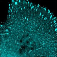
In the recent Nature paper, “Probing cellular protein complexes using single-molecule pull-down”, published May 26, 2011, researchers at the University of Illinois at Urbana-Champaign outline their findings for a new method for visualizing protein complexes through a single-molecule immunoassay which combines an antigen capturing chip and TIRF Microscopy. The SiMPull method captures protein complexes and the captured complexes are then visualized with fluorescent dyes or fluorescent protein tags. This is accomplished by using a microscope slide covered with biotinylated polyethylene glycol (PEG) and streptavidin bound to biotinylated antibodies. For single molecule visualization, multicolor labeling provides differentiation of subcomplexes and configurations.
The study includes two validation experiments, where study team members tagged their chosen complexes with YFP, in order to estimate YFP concentration after pulldown and subsequent imaging. They were then able to determine stoichiometric information in human kidney cells, from the isolated monomeric or dimeric YFPs, which exhibited the one and two-step decay responses.
Additionally, in another validation experiment, team members chose to use protein kinase A (PKA) because it’s two catalytic and two regulatory subunits separate in the presence of cAMP. This was accomplished by labeling the catalytic subunits of protein kinase A with YFP and the regulatory subunits with mCherry fluorescent protein. They then used a two-color SiMPull to pull down PKA. After pull-down they imaged the PKA in the presence of cAMP and without cAMP present. The YFP and mCherry signals fluoresced together, demonstrating that the catalytic and regulatory subunits were still attached to eachother. The YFP and mCherry signals did not correlate in the presense of cAMP, reaffirming the fact that the two subunits disassociate in the presence of cAMP.
Unlike other single-molecule pulldown techniques, the SiMPull does not require purified proteins. It also only requires about 10 cells of sample for protein pull-down and analysis, while traditional Western Blots require about 5000 cells. Moreover, This two-color SiMPull method could be further optimized yielding higher resolution overlay when used in combination with Allele’s mTFP1 and LanYFP, the brightest fluorescent proteins on the market.
The full article can be found at http://www.nature.com/nature/journal/v473/n7348/full/nature10016.html
New Product of the Week: High quality Anti-FLAG Monoclonal Antibody for detection or pulldown, ABP-MAB-DT006, $219/100ug.
Promotion of the Week: save 5% on all of our pre-packaged viruses. Our pre-packaged viruses are all titered at 10^8th or higher, and packaged in 5 tubes for your convenience. To redeem this offer email the code PREPACK to abbashussain@allelebiotech.com.


No comments:
Post a Comment