The resolution of optical microscopy is limited by the Abbe limit, the diffraction limit roughly half the wavelength of the light used (e.g. green light is around 500 nm, its Abbe limit is 250 nm). Super-imaging fluorescence often involves switching fluorophores between a dark and a bright state, building a high-resolution image from many single, localized fluorophores. These technologies have become relatively well-known in the past years, including stimulated emission depletion (STED), stochastic optical reconstruction microscopy (STORM), photoactivatable localization microscopy (PALM), and a number of variations.
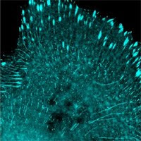
STED and saturated structured illumination (SSIM) require specialized microscopes to shrink the effective size of the scanning beam or to extract information from hidden patterns. Localization techniques require non-overlapping fluorophore emission by activating a small populations of fluorophores, e.g. those of photoswitchable FPs such as Dendra,mEos, or mClavGR2. Cox et al. recently reported in Nature Methods that by using Bayesian modeling they were able to utilize many overlapping fluorophores to obtain localization from blinking and bleaching. This allows high resolution imaging at 50 nm using wide field microscopy, regular FPs, and on live cells.
The technology’s novelty and focus were mostly on modeling. The authors used podosome (cytoskeletal structures associated with cell adhesion, migration, and disintegration of the extracellular matrix) danymics imaging as the first example to demonstrate the power of the new method. The software for data analysis is provided at http://3bmicroscopy.com. The technology is termed 3B analysis for Bayesian analysis of the Blinking and Bleaching. Bayesian: Bayesian probability as "a degree of plausibility of a proposition (belief in a proposition) based on the given state of knowledge," in contrast to interpreting it as a frequency or a "propensity" of some phenomenon.
Cox et al.: http://www.nature.com/nmeth/journal/vaop/ncurrent/full/nmeth.1812.html
Showing posts with label PALM. Show all posts
Showing posts with label PALM. Show all posts
Saturday, December 17, 2011
Thursday, January 6, 2011
New Frontiers for Research Tool Development in the New Year

Optogenetics
Chosen as the Method of the Year 2010 by Nature Method and mentioned in a number of year-end recaps, this is a technology that allows the use of light to precisely (at least in a temporal sense) control engineered proteins within a targeted cell population. For example, by introducing light-activated channelrhodopsins into neurons, one can use a pulse of light to initiate a movement of ion across the cell membrane. The technology, first reported in 2005 then made headlines as a major impact on neurosciences since 2007, is now being combined with other components in controlling a broader array of biological events, such as DNA binding, enzyme activities, etc. Looking forward, a few areas will be more than likely the frontlines of moving optogenetics into more labs:
Additional combinations: The few known channelrhodopsins and their fast growing variations will be combined with more “effecter” domains to control different events. The challenge will be to find ways to use the structural changes or any responses channelrhodopsins have to stimulating lights in order to trigger a reaction in the associated effecter domain.
Tracking mechanisms: A platter of fluorescent proteins (FPs) will be used as an independent tracking method to follow cells being targeted. FPs that have optical spectra that do not interfere with the optogenetic molecules will be tested and established. In addition, FPs with less toxicity, narrower excitation and emission peaks, and more tolerance to different cellular environment will be preferred and eventually set up as standards.
Delivery tools: To bring the optogenetic reagents into cells like neurons researchers will most likely rely on lentiviral vectors in most cases. Other vehicles such as baculovirus, MMLV-based retrovirus, even herpes virus may find broader applications in this field. Pre-packaged lentiviruses and MMLV-retroviruses already contain optogenetic constructs will become popular products.
VHH Antibodies
The small capture polypeptides based on single-domain Camelid antibodies (nanobodies) and similar VHH domains will become much dramatically more popular this year, judging from the significant increase in demands of the only camelid reagent products, GFP-Trap and RFP-Trap, in 2010. There are a number of NIH initiated programs that aim to find capture reagents that eventually target the complete human proteome. One of the key criteria for the current phase of the relevant NIH Director’s Initiative is ability to co-immunoprecipitate. The Human Proteome Organization (HUPO) recently expressed frustration due to the lack of high quality capture reagents necessary to isolate and identify most proteins. HUPO promotes global research on proteins in order to decode the human proteome. From what we have learned from dozens of publications showing the use of GFP-Trap, VHH molecules pulls down GFP-tagged proteins with unprecedented efficiency and purity. VHH antibodies show strong affinity and specificity, at a level superior or comparable to monoclonal antibodies. In addition, VHH antibodies are increasingly appreciated for their capabilities to recognize concave epitopes by their relatively convex-shaped paratopes. VHH nanobodies are small (~12-15 kD), with a limited number of functionally important disulfide bonds, can be expressed very well in E. coli, and are amazingly stable in extreme denaturing conditions such as heat and acid. They have been shown to be better suited for in vivo and trans-cellular membrane delivery than other antibodies. It should not be surprising that one day in the coming years VHH antibodies will be more dominant than monoclonal antibodies.
Super-Resolution Imaging
One of the goals of developing technologies such as photoactivated localization microscopy (PALM) and related super-resolution imaging (SRI) techniques was to achieve electron microscopy (EM) level resolution without using EM. Now new developments show that maybe combining EM and photoactivable FPs would provide more specific and more detailed morphology. It would be anticipated that more photoconvertible FPs will prove to work well for one type of SRI or another. The event that will bring this technology to nearly every cell biology lab is the improvement and availability of necessary instruments that some companies have already begun to commercialize.
New Product of the Week 010311-010911:
Human let-7b miRNA minigene on lentivirus with RFP reporter, ABP-RP-MILT7BLP
Promotion of the Week 010311-010911:
15% off mWasabi-based organelle markers carried on baculo2mammalian system if order this week (On-Demand products will require about 3-4 weeks for virus packaging after an order is placed). Use code 0103BACFP on fax or email order.
Tuesday, June 16, 2009
Allele Will Bring a New Family of Fluorescent Proteins to the Market
Allele has signed an exclusive co-development and marketing agreement with the Swedish high tech company, Innoventus, to work with Dr. Olle Israelsson of the Karolinska Institutet on a novel class of fluorescent proteins.
These proteins were discovered in Amphioxus, a type of small fish that can be found in beach sand, which is believed to be a very primitive cordate species. Compared to jellyfish and coral, from which virtually all of the currently used fluorescent proteins were isolated, Amphoixus are closer to mammalians and their proteins may find great application in human cells and other commonly used animal models. In addition, there are a large number of protein variants that can provide different spectra and other important physical properties such as photostability and photoconvertability.
Allele Biotech’s plan is to first introduce several new fluorescent proteins of different colors to the market as immediate alternatives for fluorescent protein customers. The next step is to continue to evolve and mature these proteins to create more advanced proteins with desired properties suitable for live animal imaging or more advanced applications such as PALM/STORM and SIM. Allele Biotech has on its team of fluorescent protein research staff and collaborators, some of the most highly regarded scientists. With these resources, Allele Biotech plans on committing to long-term development of truly user-friendly fluorescence imaging products.
These new class of fluorescent proteins will be integrated into Allele Biotech’s current products including: retro/lentiviral vectors, baculovirus and bacmam systems, as well as iPSC and RNAi constructs.
Subscribe to:
Posts (Atom)