This blog will be the template of Allele's new cell based assay service landing page. http://www.allelebiotech.com/allele3/index.php
Overview:
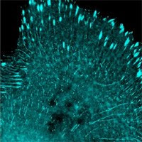
Originally cloned from the jellyfish Aequorea victoria and subsequently from many other marine organisms, fluorescent proteins (FPs) spanning the entire visual spectrum have become some of the most widely used genetically encoded tags. Unlike traditional labeling methods, FPs may be used to specifically label virtually any protein of interest in a living cell with minimal perturbation to its endogenous function. Genes encoding FPs alone or as fusions to a protein of interest may be introduced to cells by a number of different methods, including simple plasmid transfection or viral transduction. Once expressed, FPs are easily detected with standard fluorescence microscopy equipment.
Factors that should be taken into account when designing an FP-based imaging experiment include the desired wavelength(s) for detection, the pH environment of the tagged protein, the total required imaging time, and the expression level or dynamic range required for detection of promoter activity or tagged protein. Individual FPs currently available to the research community vary considerably in their photostability, pH sensitivity, and overall brightness, and so FPs must be chosen with care to maximize the likelihood of success in a particular experimental context.
FPs as fusion tags:
Use of FPs as fusion tags allows visualization of the dynamic localization of the tagged protein in living cells. For such applications, the cDNA of a protein of interest is attached in-frame to the coding sequence for the desired FP, and both are put under the control of a promoter appropriate to the experimental context (typically CMV for high-level expression, though other promoters may be desirable if overexpression of your protein of interest is suspected of producing artifacts). The most basic uses for fluorescent protein fusions include tracking of specific organelles (fusions to short organelle targeting signals) or cytoskeletal structures (fusions to actin or tubulin, for example). More advanced uses include tracking receptors or exported proteins. In most cases, it is critical that the FP used for fusion tagging be fully monomeric, as any interaction between fusion tags is likely to produce artifacts, some of which may be hard to recognize in the absence of other controls. While in most cases FP fusions do not interfere with normal protein function, whenever possible, FP fusion proteins should be validated by immunostaining the corresponding endogenous protein in non-transfected cells and verifying similar patterns of localization.
FPs as expression reporters:
FPs are highly useful as quantitative expression reporters. By driving the expression of an FP gene by a specific promoter of interest, it is possible to produce an optical readout of promoter activity. Use of the brightest possible FP ensures the best dynamic range for such an experiment. Because dynamic localization is not generally an issue for expression reporter applications, it is possible to use non-monomeric FPs for this purpose, opening up additional possibilities for multiple wavelength imaging. In order to obtain more reliable quantitative data and to correct for likely variations between individual cells in expression reporter experiments, the use of two spectrally distinct (e.g. green and red) FPs is advisable. By driving expression of one FP with a constitutive promoter and a second FP with the promoter of interest, the ratio of the two signals provides a quantitative readout of relative activity. Averaged over many cells, this technique should provide statistical power necessary for quality expression level experiments. Because FPs normally have a very slow turnover rate in mammalian cells, it may be desirable to add a degradation tag to your FP to enhance temporal resolution when measuring highly dynamic promoter activity.
Overview:
Originally cloned from the jellyfish Aequorea victoria and subsequently from many other marine organisms, fluorescent proteins (FPs) spanning the entire visual spectrum have become some of the most widely used genetically encoded tags. Unlike traditional labeling methods, FPs may be used to specifically label virtually any protein of interest in a living cell with minimal perturbation to its endogenous function. Genes encoding FPs alone or as fusions to a protein of interest may be introduced to cells by a number of different methods, including simple plasmid transfection or viral transduction. Once expressed, FPs are easily detected with standard fluorescence microscopy equipment.
Factors that should be taken into account when designing an FP-based imaging experiment include the desired wavelength(s) for detection, the pH environment of the tagged protein, the total required imaging time, and the expression level or dynamic range required for detection of promoter activity or tagged protein. Individual FPs currently available to the research community vary considerably in their photostability, pH sensitivity, and overall brightness, and so FPs must be chosen with care to maximize the likelihood of success in a particular experimental context.
FPs as fusion tags:
Use of FPs as fusion tags allows visualization of the dynamic localization of the tagged protein in living cells. For such applications, the cDNA of a protein of interest is attached in-frame to the coding sequence for the desired FP, and both are put under the control of a promoter appropriate to the experimental context (typically CMV for high-level expression, though other promoters may be desirable if overexpression of your protein of interest is suspected of producing artifacts). The most basic uses for fluorescent protein fusions include tracking of specific organelles (fusions to short organelle targeting signals) or cytoskeletal structures (fusions to actin or tubulin, for example). More advanced uses include tracking receptors or exported proteins. In most cases, it is critical that the FP used for fusion tagging be fully monomeric, as any interaction between fusion tags is likely to produce artifacts, some of which may be hard to recognize in the absence of other controls. While in most cases FP fusions do not interfere with normal protein function, whenever possible, FP fusion proteins should be validated by immunostaining the corresponding endogenous protein in non-transfected cells and verifying similar patterns of localization.
FPs as expression reporters:
FPs are highly useful as quantitative expression reporters. By driving the expression of an FP gene by a specific promoter of interest, it is possible to produce an optical readout of promoter activity. Use of the brightest possible FP ensures the best dynamic range for such an experiment. Because dynamic localization is not generally an issue for expression reporter applications, it is possible to use non-monomeric FPs for this purpose, opening up additional possibilities for multiple wavelength imaging. In order to obtain more reliable quantitative data and to correct for likely variations between individual cells in expression reporter experiments, the use of two spectrally distinct (e.g. green and red) FPs is advisable. By driving expression of one FP with a constitutive promoter and a second FP with the promoter of interest, the ratio of the two signals provides a quantitative readout of relative activity. Averaged over many cells, this technique should provide statistical power necessary for quality expression level experiments. Because FPs normally have a very slow turnover rate in mammalian cells, it may be desirable to add a degradation tag to your FP to enhance temporal resolution when measuring highly dynamic promoter activity.
New Product of the Week 03-15-10 to 03-21-10: Oct4-Sox2
2-in-1 lentivirus ABP-SC-LVI2in1 for effective iPS generation link: http://www.allelebiotech.com/shopcart/index.php?c=132&sc=122.
Promotion of the Week 03-15-10 to 03-21-10: 5% off
plate oligos at all scales! www.allelebiotech.com/allele3/Oligo_96Plate.php We are doing our “window promotion” again, during a hour-long window, get any Allele’s High efficiency competent cells at 30% regular price, the time will be announced tomorrow on our Facebook page.