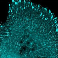
Showing posts with label FRET. Show all posts

Friday, July 30, 2010

Allele’s pallet of the super star fluorescent proteins

From AlleleBlogs
http://allelebiotech.com/blogs/2010/07/alleles-pallet-of-the-super-star-fluorescent-proteins/


“Photoblog”–just some fun pictures from our notebooks.
    The brightest cyan, green fluorescent proteins, and the brightest ever FP in LanYFP!
The brightest cyan, green fluorescent proteins, and the brightest 
ever FP in LanYFP!
Ain't they pretty?
These fluorescent proteins are representatives of the growing family or high quality, new generation FPs engineered to enable experiment previously deemed impossible.
    Cells infected with lentivirus carrying mWasabi. Lentivirus carrying LanYFP will make most cells much more brighter than this.
2-3 times brighter than EGFP, no cytotoxicity detected
The mWasabi is stimulating
The brightest green fluorescent protein with excellent photostability, carried on 10e8 TU/ml high titer lentivirus.
    The LanFPs express well in bacteria.
Reminding you of icecream
The LanFPs express well in bacteria
Project planning is under way to test the cytotoxicity of lanFPs in different mammalian cell lines and in vivo with a focus on neurons.
    The FPs fold so strongly that they fluorescence even in SDS-PAGE.
Fluorescence while running in denaturing gel
Can you see the FP bands in the SDS PAGE?
    FPs in SDS PAGE–a closer look
while the gel is still running
Can you see them now?
    FPs in gel cassette over UV lights
Easier to see now than during gel running
Invincible FPs
    FPs in gel cassette under blue LED
The red FP is harder to see because of the black background
Fluorescence in SDS page under blue LED
The purified FPs can be used as “real time” protein markers.
New Product of the Week 07/26/10-08/01/10: pCHAC-mWasabi-C for expressing mWasabi fusion through retroviral vectors.
Promotion of the Week 07/26/10-08/01/10: Get 3′ TAMRA & BHQ oligo mods for $45 ea & 3′ Dabcyl mod for $20 50 nmol syn scale only/while supplies last- use dbtkrm0726

Thursday, July 1, 2010

Allele Custom Services for Drug Screening Companies

http://allelebiotech.com/blogs/2010/06/allele-custom-services-for-drug-screening-companies/
Many target discovery and validation programs can benefit from RNA interference, fluorescent proteins, stem cells, and viral delivery systems. However, applications of these technologies require special reagents and laboratory know-how. Even when available, many generic reagent kits are not tailored for your particular needs in screening or validation.

At Allele, we accelerate your discovery efforts with custom RNAi screening, fluorescence based assays, and cell model development services.

1) Our RNAi platform, based on our patented shRNA/miRNA technologies, use DNA linear template, plasmid, lentivirus, retrovirus, or baculovirus vectors that prompt cells to endogenously express RNAi. As a result, our screens offer advantages over synthetic siRNAs:
• Higher levels of consistency
• Greater delivery and gene silencing efficiencies
• Accessibility to difficult-to-transfect cells, including primary cells
• Potential for inducible RNAi expression
• More persistent silencing with shRNA under Allele’s own IP–you may not need to license siRNA patents!

2) Fluorescent proteins (FPs), which can span the entire visual spectrum, have become some of the most widely used genetically encoded tags. Genes encoding FPs alone or as fusions to a protein of interest may be introduced to cells by a number of different methods, including simple plasmid transfection or viral transduction. Allele Biotech is one of a few companies that develop and improve FPs through fundamental research. We have so far achieved:
• The brightest cyan and green FPs, true monomers for minimum artifact or cytotoxicity
• The brightest yellow and red FPs from lancelet, only FPs from vertebrate
• mTFP1 as the best FRET donor by 3 independent reports
• Photoconvertible FPs for super imaging or kinetic labeling
• Delivery on plasmid, retrovirus, lentivirus, baculovirus vectors

3) As a major advancement in the stem cell field, it has recently been shown that mouse and human differentiated cells may be reprogrammed into stem-like, pluripotent cells by the introduction of defined transcription factors. These induced stem cells (iPSCs) provide unprecedented resources of cells of different differentiation stages for functional testing and drug screening. Allele Biotech develops and provides state-of-the-art reagents in convenient forms for iPSC production
• iPS factors carried on lentivirus, retrovirus, baculovirus for different cell types
• Availability in combination with fluorescent proteins under own IP, and drug resistant genes
• 4-in-1 or 2-in-1 effective use of iPS factors on one viral vector
• Feeder cells of human origin expressing factors essential for stem cell culturing

4) Introduction of protein factors, miRNA, promoter-reporter, and virtually any other genetic element of interest via the most efficient viral packaging systems.
• Introducing protein-FP fusion, promoter-FP reporter, photoactivatable factors for cell-based assays
• Introducing critical factors for cell immortalization
• Episomal or integrated expression using baculoviral vectors
• High throughput, systematic expression of whole class of molecules in any type of cell
• High titer viral packaging at low cost for delivery to animal tissues

In addition, the Allele team can provide custom-designed assays that can be used for assaying enzyme activities in almost any pathway, such as the EGF pathway, TNF response/apoptosis pathway, nuclear receptors, etc. We utilize technically advanced methods to provide our partners with advantages over alternative methods or other services.

New Product of the Week 06-28-10 to 07-03-10: Eco-friendly mammalian tissue culture plates, 40% less plastic to the environment, 40% less cost to your budget, contact our sales rep today for quotes and details.

Promotion of the Week 06-28-10 to 07-03-10: Oct3/4 iPS lentivirus with RFP as marker, new to the market, this week only all kits containing Oct3/4-RFP same price as the original, non-RFP versions, save ~$50!

Friday, March 19, 2010

Fluorescent Protein-Based Assay Development

This blog will be the template of Allele's new cell based assay service landing page. http://www.allelebiotech.com/allele3/index.php

Overview:

Originally cloned from the jellyfish Aequorea victoria and subsequently from many other marine organisms, fluorescent proteins (FPs) spanning the entire visual spectrum have become some of the most widely used genetically encoded tags. Unlike traditional labeling methods, FPs may be used to specifically label virtually any protein of interest in a living cell with minimal perturbation to its endogenous function. Genes encoding FPs alone or as fusions to a protein of interest may be introduced to cells by a number of different methods, including simple plasmid transfection or viral transduction. Once expressed, FPs are easily detected with standard fluorescence microscopy equipment.

Factors that should be taken into account when designing an FP-based imaging experiment include the desired wavelength(s) for detection, the pH environment of the tagged protein, the total required imaging time, and the expression level or dynamic range required for detection of promoter activity or tagged protein. Individual FPs currently available to the research community vary considerably in their photostability, pH sensitivity, and overall brightness, and so FPs must be chosen with care to maximize the likelihood of success in a particular experimental context.

FPs as fusion tags:

Use of FPs as fusion tags allows visualization of the dynamic localization of the tagged protein in living cells. For such applications, the cDNA of a protein of interest is attached in-frame to the coding sequence for the desired FP, and both are put under the control of a promoter appropriate to the experimental context (typically CMV for high-level expression, though other promoters may be desirable if overexpression of your protein of interest is suspected of producing artifacts). The most basic uses for fluorescent protein fusions include tracking of specific organelles (fusions to short organelle targeting signals) or cytoskeletal structures (fusions to actin or tubulin, for example). More advanced uses include tracking receptors or exported proteins. In most cases, it is critical that the FP used for fusion tagging be fully monomeric, as any interaction between fusion tags is likely to produce artifacts, some of which may be hard to recognize in the absence of other controls. While in most cases FP fusions do not interfere with normal protein function, whenever possible, FP fusion proteins should be validated by immunostaining the corresponding endogenous protein in non-transfected cells and verifying similar patterns of localization.

FPs as expression reporters:

FPs are highly useful as quantitative expression reporters. By driving the expression of an FP gene by a specific promoter of interest, it is possible to produce an optical readout of promoter activity. Use of the brightest possible FP ensures the best dynamic range for such an experiment. Because dynamic localization is not generally an issue for expression reporter applications, it is possible to use non-monomeric FPs for this purpose, opening up additional possibilities for multiple wavelength imaging. In order to obtain more reliable quantitative data and to correct for likely variations between individual cells in expression reporter experiments, the use of two spectrally distinct (e.g. green and red) FPs is advisable. By driving expression of one FP with a constitutive promoter and a second FP with the promoter of interest, the ratio of the two signals provides a quantitative readout of relative activity. Averaged over many cells, this technique should provide statistical power necessary for quality expression level experiments. Because FPs normally have a very slow turnover rate in mammalian cells, it may be desirable to add a degradation tag to your FP to enhance temporal resolution when measuring highly dynamic promoter activity.

Overview:

Originally cloned from the jellyfish Aequorea victoria and subsequently from many other marine organisms, fluorescent proteins (FPs) spanning the entire visual spectrum have become some of the most widely used genetically encoded tags. Unlike traditional labeling methods, FPs may be used to specifically label virtually any protein of interest in a living cell with minimal perturbation to its endogenous function. Genes encoding FPs alone or as fusions to a protein of interest may be introduced to cells by a number of different methods, including simple plasmid transfection or viral transduction. Once expressed, FPs are easily detected with standard fluorescence microscopy equipment.

Factors that should be taken into account when designing an FP-based imaging experiment include the desired wavelength(s) for detection, the pH environment of the tagged protein, the total required imaging time, and the expression level or dynamic range required for detection of promoter activity or tagged protein. Individual FPs currently available to the research community vary considerably in their photostability, pH sensitivity, and overall brightness, and so FPs must be chosen with care to maximize the likelihood of success in a particular experimental context.

FPs as fusion tags:

Use of FPs as fusion tags allows visualization of the dynamic localization of the tagged protein in living cells. For such applications, the cDNA of a protein of interest is attached in-frame to the coding sequence for the desired FP, and both are put under the control of a promoter appropriate to the experimental context (typically CMV for high-level expression, though other promoters may be desirable if overexpression of your protein of interest is suspected of producing artifacts). The most basic uses for fluorescent protein fusions include tracking of specific organelles (fusions to short organelle targeting signals) or cytoskeletal structures (fusions to actin or tubulin, for example). More advanced uses include tracking receptors or exported proteins. In most cases, it is critical that the FP used for fusion tagging be fully monomeric, as any interaction between fusion tags is likely to produce artifacts, some of which may be hard to recognize in the absence of other controls. While in most cases FP fusions do not interfere with normal protein function, whenever possible, FP fusion proteins should be validated by immunostaining the corresponding endogenous protein in non-transfected cells and verifying similar patterns of localization.

FPs as expression reporters:

FPs are highly useful as quantitative expression reporters. By driving the expression of an FP gene by a specific promoter of interest, it is possible to produce an optical readout of promoter activity. Use of the brightest possible FP ensures the best dynamic range for such an experiment. Because dynamic localization is not generally an issue for expression reporter applications, it is possible to use non-monomeric FPs for this purpose, opening up additional possibilities for multiple wavelength imaging. In order to obtain more reliable quantitative data and to correct for likely variations between individual cells in expression reporter experiments, the use of two spectrally distinct (e.g. green and red) FPs is advisable. By driving expression of one FP with a constitutive promoter and a second FP with the promoter of interest, the ratio of the two signals provides a quantitative readout of relative activity. Averaged over many cells, this technique should provide statistical power necessary for quality expression level experiments. Because FPs normally have a very slow turnover rate in mammalian cells, it may be desirable to add a degradation tag to your FP to enhance temporal resolution when measuring highly dynamic promoter activity.

New Product of the Week 03-15-10 to 03-21-10: Oct4-Sox2 2-in-1 lentivirus ABP-SC-LVI2in1 for effective iPS generation link: http://www.allelebiotech.com/shopcart/index.php?c=132&sc=122.
Promotion of the Week 03-15-10 to 03-21-10: 5% off plate oligos at all scales! www.allelebiotech.com/allele3/Oligo_96Plate.php We are doing our “window promotion” again, during a hour-long window, get any Allele’s High efficiency competent cells at 30% regular price, the time will be announced tomorrow on our Facebook page.

Wednesday, December 16, 2009

mTFP1 is an excellent FRET donor

Because of its excitation and emission wavelength, sharp excitation and emission peaks, high quantum yield, and exceptional photostability, mTFP1 has always been considered a very good Forster resonance energy transfer (FRET) donor (1). More recently, several groups have investigated the use of mTFP1 in various FRET experiments and imaging modalities and have shown that mTFP1 is indeed one of the best choices (2, 3, 4).

In one recent publication, Padilla-Parra et al (2) tested a number of different FRET couples to determine which was the best for fluorescence lifetime imaging (FLIM)-FRET experiments, and found that the mTFP1-EYFP pair was by far the best pair for FLIM-FRET. This group also confirmed that the fluorescence lifetime decay of mTFP1 fits well to a single exponential, and that the time constant for this decay is unaffected by photobleaching, making mTFP1 an excellent choice for any kind of fluorescence lifetime imaging applications, including FLIM-FRET. This group also notes that it is likely that the use of Venus or mCitrine variants in place of EYFP would improve the performance of this FRET pair even further.

In a mathematical analysis of the potential FRET efficiency of mTFP1 with Venus YFP, Day et al. (3) showed that compared with Cerulean (currently the brightest cyan Aequorea GFP variant), one can expect up to 17% better FRET efficiency using mTFP1. This group went on to characterize the mTFP1-Venus pair in live-cell FRET and FLIM-FRET experiments and showed that it worked as predicted in both cases. They also note that mTFP1 has superior brightness and photostability when compared to Cerulean in live cells, which is consistent with all in vitro data reported previously (1). In a related paper, Sun et al. (4) demonstrated that mTFP1 is also an excellent FRET donor for the orange fluorescent protein mKO2.

Together, these recent independent studies confirm that mTFP1 among the best options when choosing a fluorescent protein as a FRET donor. With its proven track record of successful fusions, mTFP1 is also an excellent all-around performer that will enhance almost any live-cell imaging experiment.

(1) Ai et al., (2006) Biochem. J. 400:531-540.
(2) Padilla-Parra et al., (2009) Biophys J. 97(8):2368-76.
(3) Day et al., (2008) J Biomed Opt. 13(3):031203.
(4) Sun et al., (2009) J Biomed Opt. 14(5):054009.

AlleleBlog Admin, by Nathan Shaner

Video of the month (NEW!): Protein Expression Systems on youtube (http://www.youtube.com/watch?v=n81orbUebsQ) and at our protein expression page.

Discount of the week (Dec 14-20): 15% off Phoenix Retrovirus Expression System 2.0 (with selection medium provided)

New product(s) of the week: 48 fluorescent protein fusions on ready-to-infect virus that get into primary mammalian cells as subcellular markers (http://www.allelebiotech.com/shopcart/index.php?c=197&sc=34), 20 infections, only $249 for a limited introduction time.