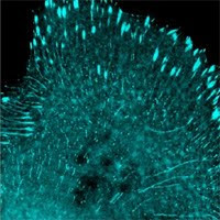
From AlleleBlog: http://blog.allelebiotech.com/2012/08/choosing-the-right-fluorescent-protein/
In 1994 the green fluorescent protein cloned from Aequorea victoria
became the first in a long line of genetically encoded labels. Since
that time, the fluorescent protein palette has expanded to cover the
entire visual spectrum. With so many color variations and options, which
fluorescent protein (FP) is best for your research? Three key factors
are among the most important to consider: brightness, photostability,
and aggregation.
Brightness is the most obvious factor that most researchers consider
when choosing an FP. In general, the brighter the FP, the better it
will perform under almost all experimental conditions. When evaluating
an FP’s brightness, make sure to look at the critical optical parameters
— extinction coefficient and quantum yield. The product of these two
values for different FPs can be used to directly compare their
brightness. Brighter FPs will have lower detection limits (i.e. the
concentration at which the FP becomes visible above autofluorescence of
other cell components), and will allow imaging with lower excitation
light intensity, minimizing the possibility of phototoxic effects.
Photostability has increasingly become a consideration when
researchers choose fluorescent proteins. Many FPs, even if they are
initially quite bright, will photobleach under continuous excitation
during imaging. In order to perform long-term imaging experiments or to
do quantitative analysis, an FP with high photostability should be the
first choice. Unfortunately, methods for measuring and reporting
photostability vary widely in the scientific literature, so be sure to
understand how your FP’s photostability was measured before trying to
make comparisons!
Aggregation (or oligomerization) has been one of the major issues
tackled in the development of FPs. Many wild-type FPs form tetramers,
which aggregate badly when expressed as fusion tags in cells. Engineered
monomeric forms of many FPs are now available, and these monomeric FPs
should always be used when making fusion constructs. For simple
expression markers, however, oligomerization is not usually a major
concern, and the brightest possible FP should be used in this case.
As with other research tools, doing your homework and reading the
primary literature is always the best approach to choosing the right FP
for your project!
Showing posts with label Fluorescent proteins. Show all posts
Showing posts with label Fluorescent proteins. Show all posts
Saturday, August 11, 2012


Saturday, December 17, 2011
Localization imaging with standard fluorescent proteins on live cells—Bayesian modeling
The resolution of optical microscopy is limited by the Abbe limit, the diffraction limit roughly half the wavelength of the light used (e.g. green light is around 500 nm, its Abbe limit is 250 nm). Super-imaging fluorescence often involves switching fluorophores between a dark and a bright state, building a high-resolution image from many single, localized fluorophores. These technologies have become relatively well-known in the past years, including stimulated emission depletion (STED), stochastic optical reconstruction microscopy (STORM), photoactivatable localization microscopy (PALM), and a number of variations.
STED and saturated structured illumination (SSIM) require specialized microscopes to shrink the effective size of the scanning beam or to extract information from hidden patterns. Localization techniques require non-overlapping fluorophore emission by activating a small populations of fluorophores, e.g. those of photoswitchable FPs such as Dendra,mEos, or mClavGR2. Cox et al. recently reported in Nature Methods that by using Bayesian modeling they were able to utilize many overlapping fluorophores to obtain localization from blinking and bleaching. This allows high resolution imaging at 50 nm using wide field microscopy, regular FPs, and on live cells.
The technology’s novelty and focus were mostly on modeling. The authors used podosome (cytoskeletal structures associated with cell adhesion, migration, and disintegration of the extracellular matrix) danymics imaging as the first example to demonstrate the power of the new method. The software for data analysis is provided at http://3bmicroscopy.com. The technology is termed 3B analysis for Bayesian analysis of the Blinking and Bleaching. Bayesian: Bayesian probability as "a degree of plausibility of a proposition (belief in a proposition) based on the given state of knowledge," in contrast to interpreting it as a frequency or a "propensity" of some phenomenon.
Cox et al.: http://www.nature.com/nmeth/journal/vaop/ncurrent/full/nmeth.1812.html
STED and saturated structured illumination (SSIM) require specialized microscopes to shrink the effective size of the scanning beam or to extract information from hidden patterns. Localization techniques require non-overlapping fluorophore emission by activating a small populations of fluorophores, e.g. those of photoswitchable FPs such as Dendra,mEos, or mClavGR2. Cox et al. recently reported in Nature Methods that by using Bayesian modeling they were able to utilize many overlapping fluorophores to obtain localization from blinking and bleaching. This allows high resolution imaging at 50 nm using wide field microscopy, regular FPs, and on live cells.
The technology’s novelty and focus were mostly on modeling. The authors used podosome (cytoskeletal structures associated with cell adhesion, migration, and disintegration of the extracellular matrix) danymics imaging as the first example to demonstrate the power of the new method. The software for data analysis is provided at http://3bmicroscopy.com. The technology is termed 3B analysis for Bayesian analysis of the Blinking and Bleaching. Bayesian: Bayesian probability as "a degree of plausibility of a proposition (belief in a proposition) based on the given state of knowledge," in contrast to interpreting it as a frequency or a "propensity" of some phenomenon.
Cox et al.: http://www.nature.com/nmeth/journal/vaop/ncurrent/full/nmeth.1812.html
Sunday, October 31, 2010
Cell Cycle Assays-Part I
From alleleblog: http://allelebiotech.com/blogs/
This is the first part of a series of blogs about using fluorescent proteins in cell based assays with established examples, a common theme here at the AlleleBlog.
FUCCI Cell Cycle Sensor
The FUCCI Cell Cycle Sensor is composed of a red (RFP) and a green (GFP) fluorescent protein fused to different regulators of the cell cycle: cdt1 and geminin.
During the cell cycle, these two proteins are ubiquitinated at different time points by specific ubiquitin E3 ligases, which tag them for degradation in the proteasome. The E3 ligases’ activities are regulated temporally and result in the biphasic cycling of GERMINI and CDT1 levels during the cell cycle. In the G1 phase of the cell cycle, GERMINI is degraded; therefore, only CDT1 tagged with RFP is present and appears as red fluorescence within the nuclei. In the S, G2, and M phases, CDT1 is degraded; only GERMINI tagged with GFP is present, resulting in cells with green fluorescent nuclei.
During the G1/S transition, when CDT1 levels are decreasing and GERMINI levels increasing, both proteins are present, so are the tagged fluorescent proteins. When the green and red images are overlaid, nuclei fluoresce yellow. This dynamic color change, from red-to-yellow-to-green, represents the entire cell cycle. This representation can be used to study the effects of elements that may influence cell cycles.
Sakaue-Sawano A, Kurokawa H, Morimura T, Hanyu A, Hama H, Osawa H, Kashiwagi S, Fukami K, Miyata T, Miyoshi H, Imamura T, Ogawa M, Masai H, Miyawaki A.Visualizing spatiotemporal dynamics of multicellular cell-cycle progression. Cell. 2008 Feb 8;132(3):487-98.CCNB1-CyclinB(NT)-GFP
In late S phage, CCNB1 promoter will be switched on to drive the expression of Cyclin B N-terminus-GFP expression; thereafter the fluorescent signal will be switched off at the destruction box in Cyclin B N-terminus at the end of Mitosis phase. During the intervening phase the fusion reporter protein will translocate from cytoplasm to nucleus by the cytoplasmic retention signal in the Cyclin B N-terminus.
Thomas N. Lighting the circle of life: fluorescent sensors for covert surveillance of the cell cycle. Cell Cycle. 2003 Nov-Dec;2(6):545-9. GFP-PCNA/YFP-PCNA
GFP-PCNA, a fusion of GFP and PCNA, has been widely used as a convenient tool to monitor the progress of S phase. At the onset of S phase, GFP-PCNA translocates into the nucleus; at mitosis the nuclear envelope breaks down and the nuclear accumulation of PCNA-GFP dissipates.
GFP-PCNA, a fusion of GFP and PCNA, has been widely used as a convenient tool to monitor the progress of S phase. At the onset of S phase, GFP-PCNA translocates into the nucleus; at mitosis the nuclear envelope breaks down and the nuclear accumulation of PCNA-GFP dissipates.
- New Product of the Week 102510-103110:
- Promotion of the Week 102510-103110:
Friday, July 30, 2010
Allele’s pallet of the super star fluorescent proteins
From AlleleBlogs
http://allelebiotech.com/blogs/2010/07/alleles-pallet-of-the-super-star-fluorescent-proteins/


http://allelebiotech.com/blogs/2010/07/alleles-pallet-of-the-super-star-fluorescent-proteins/
“Photoblog”–just some fun pictures from our notebooks.
- The brightest cyan, green fluorescent proteins, and the brightest ever FP in LanYFP!

Ain't they pretty?
These fluorescent proteins are representatives of the growing family or high quality, new generation FPs engineered to enable experiment previously deemed impossible.
- Cells infected with lentivirus carrying mWasabi. Lentivirus carrying LanYFP will make most cells much more brighter than this.

The mWasabi is stimulating
The brightest green fluorescent protein with excellent photostability, carried on 10e8 TU/ml high titer lentivirus.
- The LanFPs express well in bacteria.

The LanFPs express well in bacteria
Project planning is under way to test the cytotoxicity of lanFPs in different mammalian cell lines and in vivo with a focus on neurons.
- The FPs fold so strongly that they fluorescence even in SDS-PAGE.

Can you see the FP bands in the SDS PAGE?
- FPs in SDS PAGE–a closer look

Can you see them now?
- FPs in gel cassette over UV lights

Invincible FPs
- FPs in gel cassette under blue LED

Fluorescence in SDS page under blue LED
The purified FPs can be used as “real time” protein markers.
New Product of the Week 07/26/10-08/01/10: pCHAC-mWasabi-C for expressing mWasabi fusion through retroviral vectors.
Promotion of the Week 07/26/10-08/01/10: Get 3′ TAMRA & BHQ oligo mods for $45 ea & 3′ Dabcyl mod for $20 50 nmol syn scale only/while supplies last- use dbtkrm0726
Thursday, June 24, 2010
Brightest Ever Fluorescent Protein
http://allelebiotech.com/blogs/2010/06/brightest-ever-fluorescent-protein-2/
LanYFP, identified from lancelet (also known as amphioxus, e.g. Branchiostoma floridae), has been found to have the following properties:
Excitation 513nm
Emission 524nm
Quantum yield 0.95
Extinction coefficient 150,000
pKa ~3.5
Salt insensitive 0-500mM NaCl
LanYFP has a brightness of 143! For comparison, the brightness of the previously known brightest FPs is 95 for tdTomato, and 34 for commonly used EGFP.
Allele already has been exclusively providing the brightest cyan FP in mTFP1 (brightness of 54); and the brightest green FP in mWasabi (brightness of 56). The confirmation of LanYFP as the brightest ever FP is a major milestone of Allele’s research and development efforts in the fluorescent protein field. We are currently monomerizing LanYFP and another lancelet protein, LanRFP. Once completed, the new proteins should definitely be the FPs of choice for in vivo imaging and FRET with unprecedented utilities.
LanYFP, identified from lancelet (also known as amphioxus, e.g. Branchiostoma floridae), has been found to have the following properties:
Excitation 513nm
Emission 524nm
Quantum yield 0.95
Extinction coefficient 150,000
pKa ~3.5
Salt insensitive 0-500mM NaCl
LanYFP has a brightness of 143! For comparison, the brightness of the previously known brightest FPs is 95 for tdTomato, and 34 for commonly used EGFP.
Allele already has been exclusively providing the brightest cyan FP in mTFP1 (brightness of 54); and the brightest green FP in mWasabi (brightness of 56). The confirmation of LanYFP as the brightest ever FP is a major milestone of Allele’s research and development efforts in the fluorescent protein field. We are currently monomerizing LanYFP and another lancelet protein, LanRFP. Once completed, the new proteins should definitely be the FPs of choice for in vivo imaging and FRET with unprecedented utilities.
Wednesday, December 16, 2009
mTFP1 is an excellent FRET donor
Because of its excitation and emission wavelength, sharp excitation and emission peaks, high quantum yield, and exceptional photostability, mTFP1 has always been considered a very good Forster resonance energy transfer (FRET) donor (1). More recently, several groups have investigated the use of mTFP1 in various FRET experiments and imaging modalities and have shown that mTFP1 is indeed one of the best choices (2, 3, 4).
In one recent publication, Padilla-Parra et al (2) tested a number of different FRET couples to determine which was the best for fluorescence lifetime imaging (FLIM)-FRET experiments, and found that the mTFP1-EYFP pair was by far the best pair for FLIM-FRET. This group also confirmed that the fluorescence lifetime decay of mTFP1 fits well to a single exponential, and that the time constant for this decay is unaffected by photobleaching, making mTFP1 an excellent choice for any kind of fluorescence lifetime imaging applications, including FLIM-FRET. This group also notes that it is likely that the use of Venus or mCitrine variants in place of EYFP would improve the performance of this FRET pair even further.
In a mathematical analysis of the potential FRET efficiency of mTFP1 with Venus YFP, Day et al. (3) showed that compared with Cerulean (currently the brightest cyan Aequorea GFP variant), one can expect up to 17% better FRET efficiency using mTFP1. This group went on to characterize the mTFP1-Venus pair in live-cell FRET and FLIM-FRET experiments and showed that it worked as predicted in both cases. They also note that mTFP1 has superior brightness and photostability when compared to Cerulean in live cells, which is consistent with all in vitro data reported previously (1). In a related paper, Sun et al. (4) demonstrated that mTFP1 is also an excellent FRET donor for the orange fluorescent protein mKO2.
Together, these recent independent studies confirm that mTFP1 among the best options when choosing a fluorescent protein as a FRET donor. With its proven track record of successful fusions, mTFP1 is also an excellent all-around performer that will enhance almost any live-cell imaging experiment.
(1) Ai et al., (2006) Biochem. J. 400:531-540.
(2) Padilla-Parra et al., (2009) Biophys J. 97(8):2368-76.
(3) Day et al., (2008) J Biomed Opt. 13(3):031203.
(4) Sun et al., (2009) J Biomed Opt. 14(5):054009.
AlleleBlog Admin, by Nathan Shaner
Video of the month (NEW!): Protein Expression Systems on youtube (http://www.youtube.com/watch?v=n81orbUebsQ) and at our protein expression page.
Discount of the week (Dec 14-20): 15% off Phoenix Retrovirus Expression System 2.0 (with selection medium provided)
New product(s) of the week: 48 fluorescent protein fusions on ready-to-infect virus that get into primary mammalian cells as subcellular markers (http://www.allelebiotech.com/shopcart/index.php?c=197&sc=34), 20 infections, only $249 for a limited introduction time.
In one recent publication, Padilla-Parra et al (2) tested a number of different FRET couples to determine which was the best for fluorescence lifetime imaging (FLIM)-FRET experiments, and found that the mTFP1-EYFP pair was by far the best pair for FLIM-FRET. This group also confirmed that the fluorescence lifetime decay of mTFP1 fits well to a single exponential, and that the time constant for this decay is unaffected by photobleaching, making mTFP1 an excellent choice for any kind of fluorescence lifetime imaging applications, including FLIM-FRET. This group also notes that it is likely that the use of Venus or mCitrine variants in place of EYFP would improve the performance of this FRET pair even further.
In a mathematical analysis of the potential FRET efficiency of mTFP1 with Venus YFP, Day et al. (3) showed that compared with Cerulean (currently the brightest cyan Aequorea GFP variant), one can expect up to 17% better FRET efficiency using mTFP1. This group went on to characterize the mTFP1-Venus pair in live-cell FRET and FLIM-FRET experiments and showed that it worked as predicted in both cases. They also note that mTFP1 has superior brightness and photostability when compared to Cerulean in live cells, which is consistent with all in vitro data reported previously (1). In a related paper, Sun et al. (4) demonstrated that mTFP1 is also an excellent FRET donor for the orange fluorescent protein mKO2.
Together, these recent independent studies confirm that mTFP1 among the best options when choosing a fluorescent protein as a FRET donor. With its proven track record of successful fusions, mTFP1 is also an excellent all-around performer that will enhance almost any live-cell imaging experiment.
(1) Ai et al., (2006) Biochem. J. 400:531-540.
(2) Padilla-Parra et al., (2009) Biophys J. 97(8):2368-76.
(3) Day et al., (2008) J Biomed Opt. 13(3):031203.
(4) Sun et al., (2009) J Biomed Opt. 14(5):054009.
AlleleBlog Admin, by Nathan Shaner
Video of the month (NEW!): Protein Expression Systems on youtube (http://www.youtube.com/watch?v=n81orbUebsQ) and at our protein expression page.
Discount of the week (Dec 14-20): 15% off Phoenix Retrovirus Expression System 2.0 (with selection medium provided)
New product(s) of the week: 48 fluorescent protein fusions on ready-to-infect virus that get into primary mammalian cells as subcellular markers (http://www.allelebiotech.com/shopcart/index.php?c=197&sc=34), 20 infections, only $249 for a limited introduction time.
Friday, December 4, 2009
Construction of An Image Library
The American Society for Cell Biology (ASCB) is “pleased to announce the receipt of a U.S. National Institutes of Health Grand Opportunities (GO) grant to build The Cell: An Image Library. The ASCB will be hiring eight cell biologists or microscopists, each at 25% time,” The job description includes, according to an email job posting, “selecting exemplary images and videos and providing metadata for short tags or descriptions as well as longer annotations including technical details crucial for image interpretations. Annotators will select related key words and note biological source, context, item type, etc., in accordance with set guidelines. Annotators will upload images and videos to the Society’s new image library for research and education.” The grant is in the million dollar range.
The need for creating an extensive image library is deservingly recognized by this “GO” grant from the stimulus program awarded to the NIH by the federal government. The difficult part will be to maintain such an image center once the grant runs out. Will it be kept up-to-date and relevant, or left to collect dust on the old images? We wish that the program would be a great success and that the NIH money well spent.
Allele Biotech has applied to the same round of NIH grants with a related proposal that, rather than cell images in general, focuses more on cell differentiation/dedifferentiation through the use of iPS cells. Title: Foundation for “Subcellular Structureome” as Stem Cell Differentiation Parameters. Summary: The key question to be addressed is how to characterize differentiating stem cells along different lineages definitively and continuously, without disrupting or disturbing the differentiating cells. The broad and long-term goals are to find ways of describing stem cell differentiation in more detailed steps, thereby providing methods to predict and direct cell fate commitment.
Aim 1 Create a panel of cells that can be reprogrammed into induced pluripotent stem cells (iPSCs) with fluorescent protein (FP) fusion markers for each organelle
.Human fibroblasts and keratinocytes will be selected from a large collection of primary human cells, based on their ease to grow and transfect, number of potential cell passages, and potentials for reprogramming with induction reagents. A group of 24 subcellular localization polypeptides (LP) and FP fusion protein constructs currently offered by Allele Biotech will be stably transfected into the selected cell.
Aim 2 Characterize the morphological changes of subcellular structures during iPSC differentiation.
Transfected primary cells that stably express subcellular localization marker proteins will be induced with either current retroviral/lentiviral vectors based reprogramming cDNAs, or a non-integrating baculoviral vector under development at Allele Biotech. These cells, 48 lines in total, will be maintained and expanded under stem cell culture conditions, then induced to differentiate into chondracytes or keratinocytes as examples of cell fate. Morphology data will be analyzed and recorded at each known stage and additional “substage” to be defined in the process.
Aim 3 Correlate morphological changes to known molecular properties of each stage and provide a “signature” set of morphological changes for each stage of each lineage
Signature morphological changes, i.e. significantly different shape, location, sub-type, and copies of organelles in a cell compared to its immediate upstream stage, will be correlated to results obtained by standard expression assays at the RNA and protein levels.
Aim 4 Use the morphology parameters to establish more defined stages of cell fate commitments
Data points will be used to create a novel morphology-based cell fate commitment atlas, which will be very helpful in guiding the stem cell and regenerate medicine research at molecular biology, cell biology and physiology levels.
Aim 5 Construct more FP fusions as organelle-specific markers and combine with stage specific gene promoter driven markers
If necessary, we plan to identify more LPs as fusion marker partners after obtaining the initial set of data, and to expand the signature morphology image database. The database can be further complemented with stage-specific gene promoter driven FP images.
Weekly Promotion of Nov 30-Dec 6: 15% off luciferase assay kit ABP-PA-ABLA011 1000 reactions at only $250.00 212.50. Compare it to what you normally pay for firefly luciferase assays and find out how much you are saving.
Reminder: Allele Biotech Spotlight Promo for ASCB Dec 09 Meeting is still on, order by Dec 9th on iPS and FP groups!
New Product of the Week of Nov 30-Dec 6: Allele Biotech’s ProperFold expression vector with fluorescent protein as indicator for proper protein folding, tracking, and purification. pORB-mWasabi+-sIRES-VSVG
The need for creating an extensive image library is deservingly recognized by this “GO” grant from the stimulus program awarded to the NIH by the federal government. The difficult part will be to maintain such an image center once the grant runs out. Will it be kept up-to-date and relevant, or left to collect dust on the old images? We wish that the program would be a great success and that the NIH money well spent.
Allele Biotech has applied to the same round of NIH grants with a related proposal that, rather than cell images in general, focuses more on cell differentiation/dedifferentiation through the use of iPS cells. Title: Foundation for “Subcellular Structureome” as Stem Cell Differentiation Parameters. Summary: The key question to be addressed is how to characterize differentiating stem cells along different lineages definitively and continuously, without disrupting or disturbing the differentiating cells. The broad and long-term goals are to find ways of describing stem cell differentiation in more detailed steps, thereby providing methods to predict and direct cell fate commitment.
Aim 1 Create a panel of cells that can be reprogrammed into induced pluripotent stem cells (iPSCs) with fluorescent protein (FP) fusion markers for each organelle
.Human fibroblasts and keratinocytes will be selected from a large collection of primary human cells, based on their ease to grow and transfect, number of potential cell passages, and potentials for reprogramming with induction reagents. A group of 24 subcellular localization polypeptides (LP) and FP fusion protein constructs currently offered by Allele Biotech will be stably transfected into the selected cell.
Aim 2 Characterize the morphological changes of subcellular structures during iPSC differentiation.
Transfected primary cells that stably express subcellular localization marker proteins will be induced with either current retroviral/lentiviral vectors based reprogramming cDNAs, or a non-integrating baculoviral vector under development at Allele Biotech. These cells, 48 lines in total, will be maintained and expanded under stem cell culture conditions, then induced to differentiate into chondracytes or keratinocytes as examples of cell fate. Morphology data will be analyzed and recorded at each known stage and additional “substage” to be defined in the process.
Aim 3 Correlate morphological changes to known molecular properties of each stage and provide a “signature” set of morphological changes for each stage of each lineage
Signature morphological changes, i.e. significantly different shape, location, sub-type, and copies of organelles in a cell compared to its immediate upstream stage, will be correlated to results obtained by standard expression assays at the RNA and protein levels.
Aim 4 Use the morphology parameters to establish more defined stages of cell fate commitments
Data points will be used to create a novel morphology-based cell fate commitment atlas, which will be very helpful in guiding the stem cell and regenerate medicine research at molecular biology, cell biology and physiology levels.
Aim 5 Construct more FP fusions as organelle-specific markers and combine with stage specific gene promoter driven markers
If necessary, we plan to identify more LPs as fusion marker partners after obtaining the initial set of data, and to expand the signature morphology image database. The database can be further complemented with stage-specific gene promoter driven FP images.
Weekly Promotion of Nov 30-Dec 6: 15% off luciferase assay kit ABP-PA-ABLA011 1000 reactions at only $
Reminder: Allele Biotech Spotlight Promo for ASCB Dec 09 Meeting is still on, order by Dec 9th on iPS and FP groups!
New Product of the Week of Nov 30-Dec 6: Allele Biotech’s ProperFold expression vector with fluorescent protein as indicator for proper protein folding, tracking, and purification. pORB-mWasabi+-sIRES-VSVG
Thursday, July 30, 2009
Why Allele?
Allele provides you with tools that you will find very helpful. The two main motives for Allele developed products are:
1) To incorporate the most advanced technologies in the field
2) To provide equal utility as other companies’ equivalent products at a much more reasonable cost.
How did we do it? By developing technologies internally, in most cases with government grant funding, by in-licensing others’ discoveries, and by listening to you, our customers.
What else do we do? Conduct basic curiosity-driven research just like most of our customers. It helps to stay on the edge and connect to the community.
1) To incorporate the most advanced technologies in the field
2) To provide equal utility as other companies’ equivalent products at a much more reasonable cost.
How did we do it? By developing technologies internally, in most cases with government grant funding, by in-licensing others’ discoveries, and by listening to you, our customers.
What else do we do? Conduct basic curiosity-driven research just like most of our customers. It helps to stay on the edge and connect to the community.
Subscribe to:
Posts (Atom)

 Posts
Posts